
- #Hippocampus anatomy in split brain full#
- #Hippocampus anatomy in split brain plus#
- #Hippocampus anatomy in split brain series#
The subiculum is the final stage in the pathway, combining information from the CA1 projection and EC layer III to also send information along the output pathways of the hippocampus. In turn, CA1 projects to the subiculum as well as sending information along the aforementioned output paths of the hippocampus. Region CA1 receives input from the CA3 subfield, EC layer III and the nucleus reuniens of the thalamus (which project only to the terminal apical dendritic tufts in the stratum lacunosum-moleculare). Region CA3 combines this input with signals from EC layer II and sends extensive connections within the region and also sends connections to region CA1 through a set of fibers called the Schaffer collaterals.

Perforant path input from EC layer II enters the dentate gyrus and is relayed to region CA3 (and to mossy cells, located in the hilus of the dentate gyrus, which then send information to distant portions of the dentate gyrus where the cycle is repeated). The lamellar concept is still sometimes considered to be a useful organizing principle, but more recent data, showing extensive longitudinal connections within the hippocampal system, have required it to be substantially modified.
#Hippocampus anatomy in split brain series#
This observation was the basis of his lamellar hypothesis, which proposed that the hippocampus can be thought of as a series of parallel strips, operating in a functionally independent way. The perforant path-to-dentate gyrus-to-CA3-to-CA1 was called the trisynaptic circuit by Per Andersen, who noted that thin slices could be cut out of the hippocampus perpendicular to its long axis, in a way that preserves all of these connections. Subicular neurons send their axons mainly to the EC. Pyramidal cells of CA1 send their axons to the subiculum and deep layers of the EC. Pyramidal cells of CA3 send their axons to CA1. Granule cells of the DG send their axons (called "mossy fibers") to CA3. There is also a distinct pathway from layer 3 of the EC directly to CA1. These axons arise from layer 2 of the entorhinal cortex (EC), and terminate in the dentate gyrus and CA3. Most external input comes from the adjoining entorhinal cortex, via the axons of the so-called perforant path. The major pathways of signal flow through the hippocampus combine to form a loop.
#Hippocampus anatomy in split brain plus#
Most anatomists use the term "hippocampus proper" to refer to the four CA fields, and "hippocampal formation" to refer to the hippocampus proper plus dentate gyrus and subiculum.

After this comes a pair of ill-defined areas called the presubiculum and parasubiculum, then a transition to the cortex proper (mostly the entorhinal area of the cortex). After CA1 comes an area called the subiculum. The CA areas are all filled with densely packed pyramidal cells similar to those found in the neocortex. Next come a series of Cornu Ammonis areas: first CA4 (which underlies the dentate gyrus), then CA3, then a very small zone called CA2, then CA1. The first of these, the dentate gyrus (DG), is actually a separate structure, a tightly packed layer of small granule cells wrapped around the end of the hippocampus proper, forming a pointed wedge in some cross-sections, a semicircle in others. Starting at the dentate gyrus and working inward along the S-curve of the hippocampus means traversing a series of narrow zones. Neuroimaging pictures can show a number of different shapes, depending on the angle and location of the cut.īasic circuit of the hippocampus, shown using a modified drawing by Ramon y Cajal. Due to the three-dimensional curvature of this structure, two-dimensional sections such as shown are commonly seen. In primate brains, including humans, the portion of the hippocampus near the base of the temporal lobe is much broader than the part at the top. For example, in the rat, the two hippocampi look similar to a pair of bananas, joined at the stems.
#Hippocampus anatomy in split brain full#
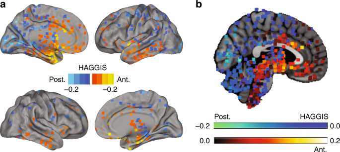
This general layout holds across the full range of mammalian species, from hedgehog to human, although the details vary. Hippocampus anatomy describes the physical aspects and properties of the hippocampus, a neural structure in the medial temporal lobe of the brain that has a distinctive, curved shape that has been likened to the sea horse monster of Greek mythology and the ram's horns of Amun in Egyptian mythology. Nissl-stained coronal section of the brain of a macaque monkey, showing hippocampus (circled).


 0 kommentar(er)
